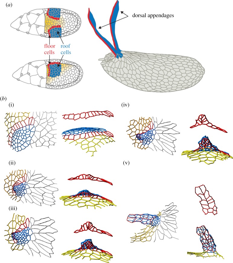
Figure 4.
Dorsal appendage formation during Drosophila oogenesis. (a) Schematic of cell types in the developing Drosophila egg chamber and the fully formed eggshell. Blue highlights the roof cells, red the floor cells, yellow the midline cells and rest are main body cells. (bi–bv) Three-dimensional reconstructions of the apical surface of follicle cells at different time points during dorsal appendage formation in D. melanogaster. The flat primordium is transformed into a conical structure through a sequence of cell neighbour exchanges. The colour-coding of the cell contours in (b) is same as the colouring of the cell surface in (a). Images are taken from [39].

