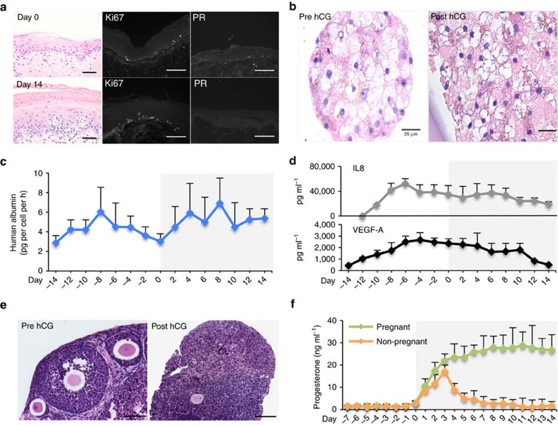
Figure 6. The Quintet-MFP supported female reproductive and non-reproductive tissues and pregnancy-like hormone condition.
(a) Ectocervix histology, and Ki67 and PR staining at the end of follicular (day 0) and luteal (day 14) phases. (b) Histology of liver microtissues before (day −14) and after (day 14) hCG treatment. (c) Human albumin production over 28 days of microfluidic culture. (d) Production of IL8 and VEGF over 28 days of microfluidic culture. (e) Histology of ovarian tissue in the Quintet-MFP on day 0 (pre hCG) and day 8 (continuously cultured with hCG). (f) Ovarian progesterone secretion with and without hCG treatment. Graphs in c,d,f display average+s.d.'. Scale bar, 10 μm (a), 25 μm (b), and 100 μm (e). CL, corpus luteum; IL8, interleukin 8; VEGF-A vascular endothelial growth factor A. n=3 replicates of the integrated tissue culture in the Quintet-MFP.

