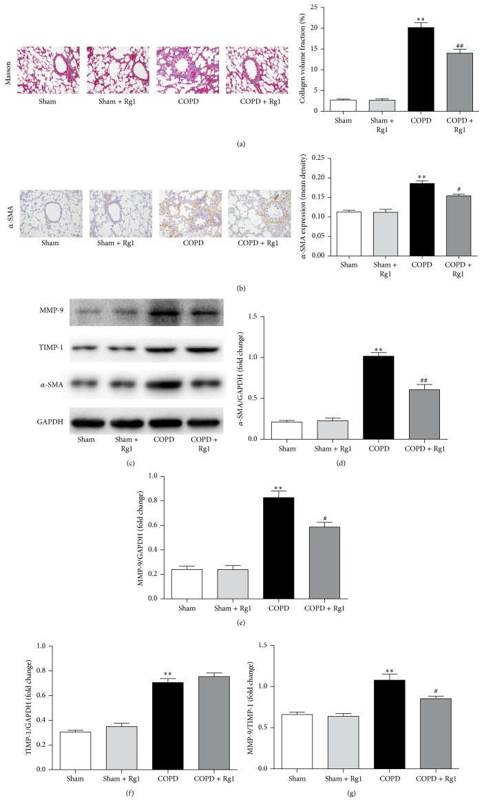
Figure 2.
Effects of ginsenoside Rg1 on airway fibrosis in chronic obstructive pulmonary disease (COPD) rats. (a) Masson trichrome staining of lung tissues. Scale bar = 100 μm. All fields shown are representative of at least six fields observed in four rats for each group. Quantitative collagen and elastin fiber assay was performed using Image-Pro Plus 6.0 software. (b) Immunohistochemical staining of α-SMA in lung tissues. Scale bar = 100 μm. All fields shown are representative of at least six fields observed in four rats for each group. Quantification of α-SMA was carried out using Image-Pro Plus 6.0 software. (c) Protein expression of α-SMA, MMP-1, and TIMP-1 in lung tissues was determined by western blot analysis. (d–f) Quantitative analysis of α-SMA, MMP-9, and TIMP-1 protein expression. (g) Ratio of MMP-9 to TIMP-1. Data are shown as mean ± SD of at least three independent experiments, each performed in triplicate. ∗∗p < 0.01 versus the Sham group; #p < 0.05 and ##p < 0.01 versus the COPD group.

