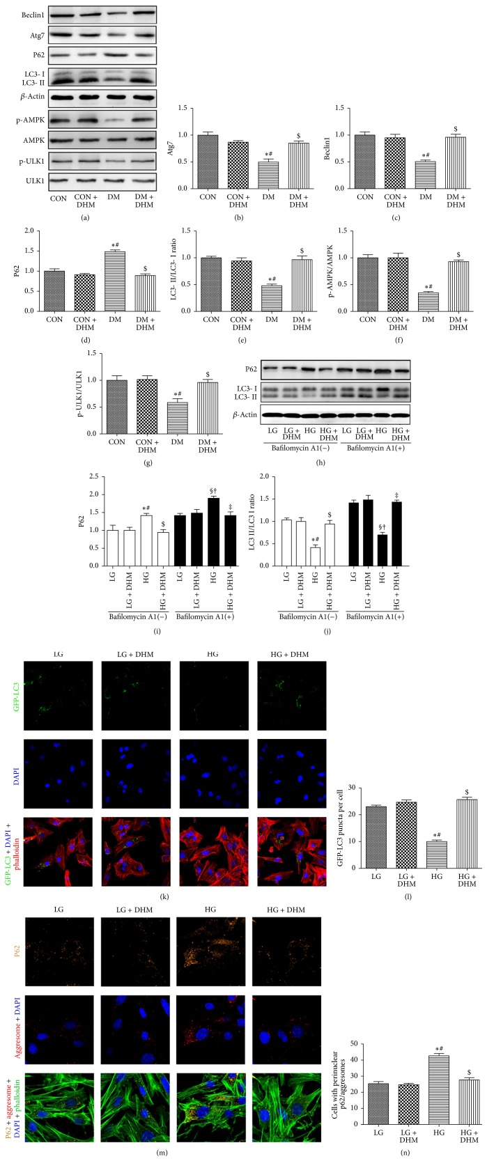
Figure 6.
Effects of DHM on cardiac autophagy in STZ-induced diabetic mice. (a)–(g) Representative immunoblots for Beclin1, Atg7, P62, LC3 II, p-AMPK, AMPK, p-ULK1, ULK1, and β-actin in myocardial tissues from the respective groups and densitometric quantification. The columns and error bars represent the means and SD (n = 6). ∗P < 0.05 versus CON; #P < 0.05 versus CON + DHM; $P < 0.05 versus DM. (h)–(j) Representative blots and analysis of P62 and LC3 in the absence or presence of bafilomycin A1. (k) DHM increased the number of GFP-LC3 puncta in high glucose-cultured neonatal cardiomyocytes. (l) Quantitative analysis of the number of GFP-LC3 puncta. (m) DHM decreased the accumulation of P62 (orange) and aggresomes (red) in high glucose-cultured neonatal cardiomyocytes. (n) Quantitative analysis of the mean numbers of autophagosomes and autolysosomes. The columns and error bars represent the means and SD. ∗P < 0.05 versus LG; #P < 0.05 versus LG + DHM; $P < 0.05 versus HG; §P < 0.05 versus LG + bafilomycin A1; †P < 0.05 versus LG + DHM + bafilomycin A1; ‡P < 0.05 versus HG + bafilomycin A1.

