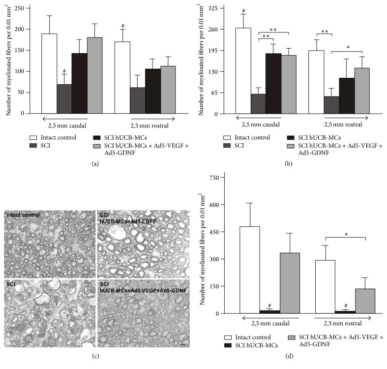
Figure 2.
Analysis of myelinated fibers. The average number of spared myelinated fibers in the lateral (a) and ventral funiculi (b) of spinal segments 2.5 mm rostral and caudal to the lesion epicenter at day 30 after injury. Differences were statistically significant between SCI/intact and other experimental groups (#P < 0.05). ∗P < 0.05, ∗∗P < 0.01 one-way ANOVA followed by a Tukey's post hoc test. (с) Fragments of the ventral funiculi at a distance of 2.5 mm from the SCI/Th8 epicenter in the caudal direction at day 30 after injury. The images are methylene blue-stained semithin sections. Scale bar: 10 μm. (d) The average number of spared myelinated fibers in the CST of spinal segments 2.5 mm caudal and rostral to the lesion epicenter/Th8 at day 30 after injury. Differences were statistically significant between SCI hUCB-MCs and other experimental groups (#P < 0.01). ∗P < 0.05 one-way ANOVA followed by a Tukey's post hoc test.

