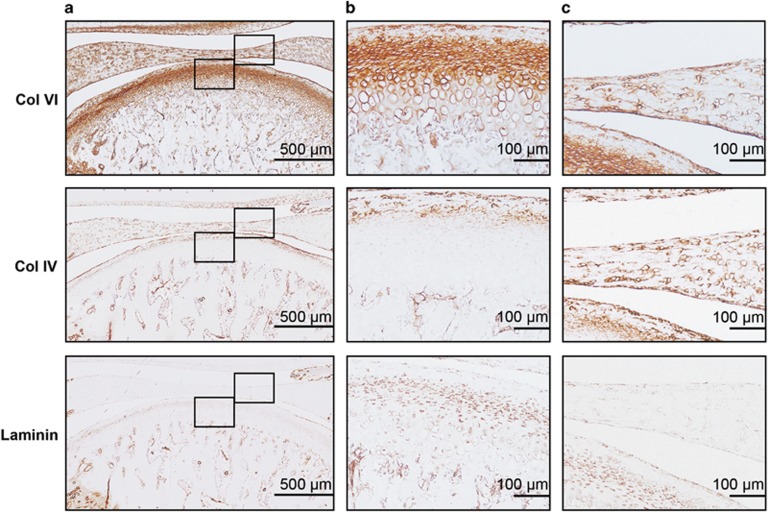
Figure 2.
Expression of PCM molecules in rat TMJ. (a) Histological sections of rat TMJ stained for collagen VI, collagen IV and laminin. Scale bars, 500 μm. (b) High magnification view of condylar cartilage showing the pericellular staining of collagen VI, collagen IV and laminin. Scale bars, 100 μm. (c) High magnification view of disc showing the pericellular staining of collagen VI, collagen IV and laminin. Scale bars, 100 μm. Representative images; n=6. PCM, pericellular matrix; TMJ, temporomandibular joint.

