Figure 3.
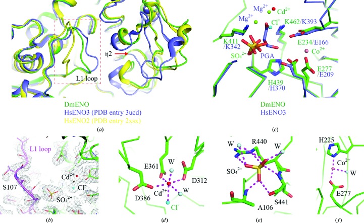
(a) Structural comparison of DmENO with HsENO2 and HsENO3. DmENO, HsENO2 and HsENO3 are coloured green, slate and yellow, respectively. The red box indicates the L1 loop region. The red arrow shows that the η2 helix is distant from the active site. (b) The 2mF o − DF c map (grey mesh) for the active site of DmENO contoured at 1.0σ. The L1 loop is coloured purple. (c) Structural comparison of the active sites of DmENO and HsENO3. HsENO3 possesses two magnesium ions and the substrate PGA in its active site, while DmENO has one cadmium ion, one cobalt ion, one chloride ion and one sulfate ion in the active site. Residues involved in substrate binding and catalysis in DmENO and HsENO3 are shown as sticks. (d) The geometry and coordinates of the cadmium ion in the active site of DmENO. (e) The residues and waters interacting with the sulfate ion in the active site of DmENO. (f) The coordination of the cobalt ion in the active site of DmENO. The magenta dashed lines represent salt bridges and hydrogen bonds.

