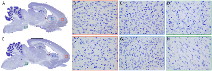
Figure 3. Absence of aggregated α-synuclein in Octodon degus brains.
(A,E) Sagittal brain sections of young (A) and aged (E) octodons showing the distribution of proteinase-K (PK) resistant α-synuclein (α-syn) immunostaining. Representative pictures of PK-resistant α-syn staining in 3 brain regions: cortex (B,F), dorsal striatum (C,G), and substantia nigra (D,H). Scale bar: 100 μm.

