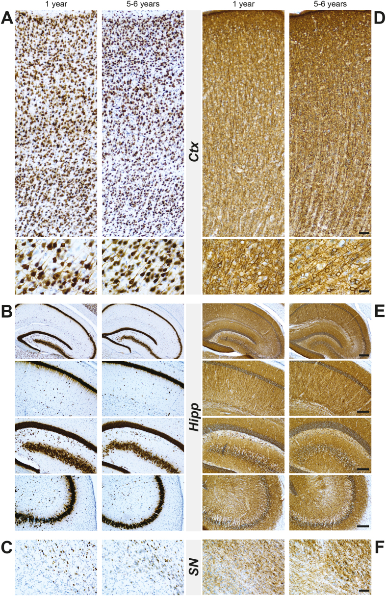
Figure 6. Profile of NeuN and Map2 immunostaining in the cortex, hippocampus and substantia nigra in wild-type young (1-year-old) and aged (5–6 years old) octodons.
(A–C) Show representative cortical (A), hippocampal (B), nigral (C) images with low and high magnifications in young and aged octodons without clear changes in average density of neurons. (D–F) Show representative images of Map2 immunostaining in cortical (D), hippocampal (E), and nigral (F) areas from young and aged octodons. Scale bar (from the top to the bottom): (A–D) 40 μm; 20 μm; (B–E) 200 μm; 100 μm; 100 μm; 100 μm; (C–F) 40 μm.

