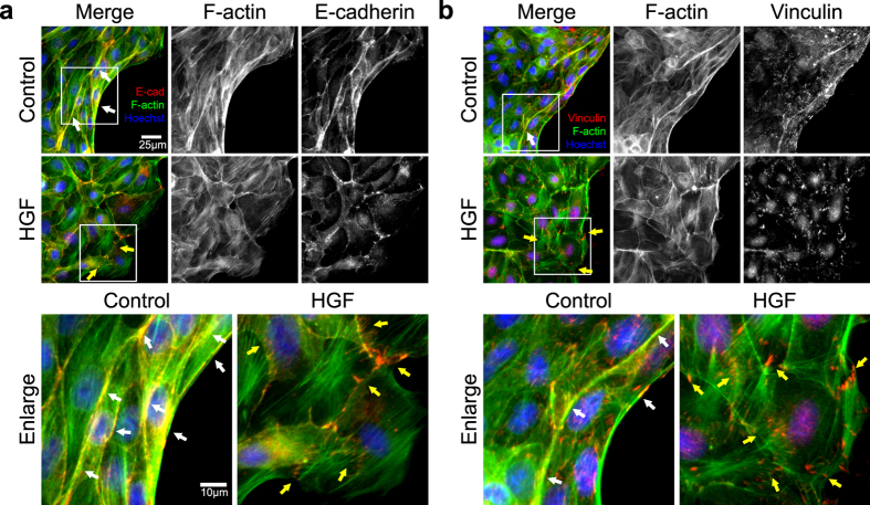
Figure 4. Immunofluorescence assays on tension-related machinery proteins in the cellular islands.
To validate the interference of co-localization by HGF, an immunofluorescence assay was performed on E-cadherin and vinculin in the islands that were fixed 9 hours after removing PDMS mask. F-actin and cell nuclei were also fluorescently labeled. The white boxes in the upper panels are enlarged at lower panels. (a) E-cadherin was expressed as distinct and thick lines with co-localization with F-actin bundles in the control sample (white arrows) but was expressed as fuzzy and punctate zones with dissociated-F-actin in HGF sample (yellow arrows). (Red; E-cadherin, green; F-actin, and blue; Hoechst) (b) Vinculin was expressed as thin lines along with F-actin bundles (white arrows) and hazy dots linked at the ends of F-actin in the control sample but was expressed as dispersed and distinct dots along the individual cell boundaries in HGF samples (yellow arrows). (Red; vinculin, green; F-actin, and blue; Hoechst).

