
Figure 5. Elastic fibre content of ovine vaginal wall.
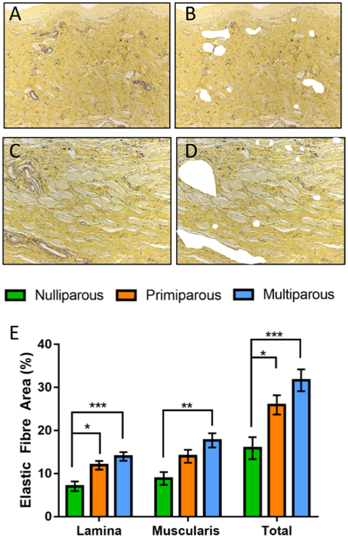
Shown as black fibres in Hart’s-stained tissue in representative images of multiparous (A) lamina propria with (B) blood vessels removed from analysis and in the (C) muscularis with (D) blood vessels removed from analysis. (E) Graph showing the image analysis quantification of elastic fibre area in tissue images with blood vessels electronically removed. n = 6, primiparous and multiparous n = 8 vaginal tissues/group. Data are mean +/− SEM, *p < 0.05, **p < 0.01, ***p < 0.001.