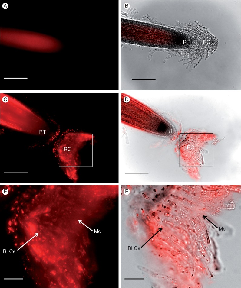
Fig. 6.
Immunolabelling of the root cap and associated border-like cells (BLCs) of Heliophila coronopifolia with the anti-Hc-AFP2 polyclonal antibody using a fluorescent secondary antibody, after pre-treatment with pectinase. (A, B) Controls with a mammalian primary antibody (non-plant negative control) (p-p38, see Table S2 for details on antibodies). (C, D) The root tip with its BLCs and (E, F) BLCs at higher magnification. Images with immunofluorescence mode (A, C, E), and images with overlayed transmission and immunofluorescence mode (B, D, F). Arrows show specific labelling with primary antibody. RT, root tip; RC, root cap; BLCs, border-like cells; Mc, mucilage. Scale bars: 200 μm (A–D), 400 μm (E, F).

