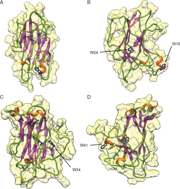
Fig. 6.
Molecular modelling of Nictaba and an NLL protein from soybean. Ribbon diagrams show front (A, C) and side (C, D) view of Nictaba and GmNLL1, respectively. The molecular surface, α-helices, β-sheets and loop/turns are coloured yellow, orange, purple and green, respectively. The conserved tryptophan residues important for the carbohydrate-binding activity of Nictaba (w15, w24, w34, w41) are indicated.

