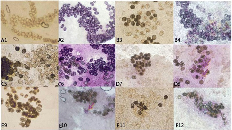
Fig. 2.

SDHG staining results for S. japonicum eggs in mouse colonic tissue. Eggs deposited in colon tissues of mice at 45 days post infection (PI) were light yellow when unstained (A1) and positive eggs ranged from light to dark purple after MTT staining (A2). Eggs deposited in colon tissues of mice 90 days PI were light yellow, light black and black when unstained (B3) and after staining with MTT, most eggs appeared light purple or purple (B4). Eggs deposited in colon tissues of mice at 180 days PI were light yellow, brownish, light black or black when unstained (C5) and following staining with MTT most eggs revealed a positive purple response while a minority were not stained, a negative response (C6). Eggs deposited in colon tissues from mice at 30 days post PZQ treatment were light black and black with a few yellow eggs when unstained (D7); after staining with MTT few eggs were light purple (red arrows), indicating a weak positive reaction (D8). Ninety days PT eggs deposited in unstained mouse colon tissue were only light black and black (E9); after staining with MTT only a few eggs were light purple (red arrow) indicating a weak positive reaction (E10). A hundred and eighty days PT unstained eggs in mouse colon tissue were either empty (yellow) or black (F11) and after staining with MTT no eggs showed a positive color reaction (F12). All images are at ×100 magnification
