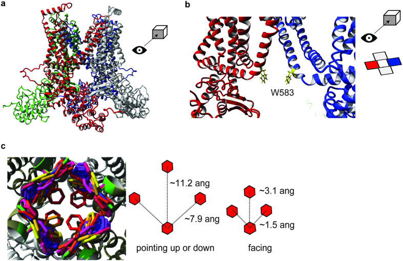
Figure 3. Homology model of TRPV5.
(a) The tetrameric TRPV5 structure is depicted in front view. Each monomer is color-coded. (b) The detailed front view of the pore region highlights the side chains of W583 (yellow) in two monomers. The side chains are sticking towards the permeation pathway. (c) The possible rotameric positions for the side chain of W583 are shown in different colors (left panel). Overall, three main rotameric positions are detected with the side chains either pointing upwards, downwards or pointing towards each other. The distance between the W583 side chains is based on the homology model and is depicted for the main rotameric positions (right panel).

