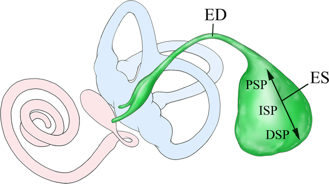
Figure 1. Schematic illustration of the membranous labyrinth of the inner ear represents the endolymphatic duct (ED; green) and sac (ES; green) and its relationship to the cochlea (red) and the vestibule (utricle and semicircular canals; blue).

The magnified view shows the ES three parts, from anterior to posterior: the proximal sac portion (PSP), the intermediate sac portion (ISP) and the distal sac portion (DSP).
