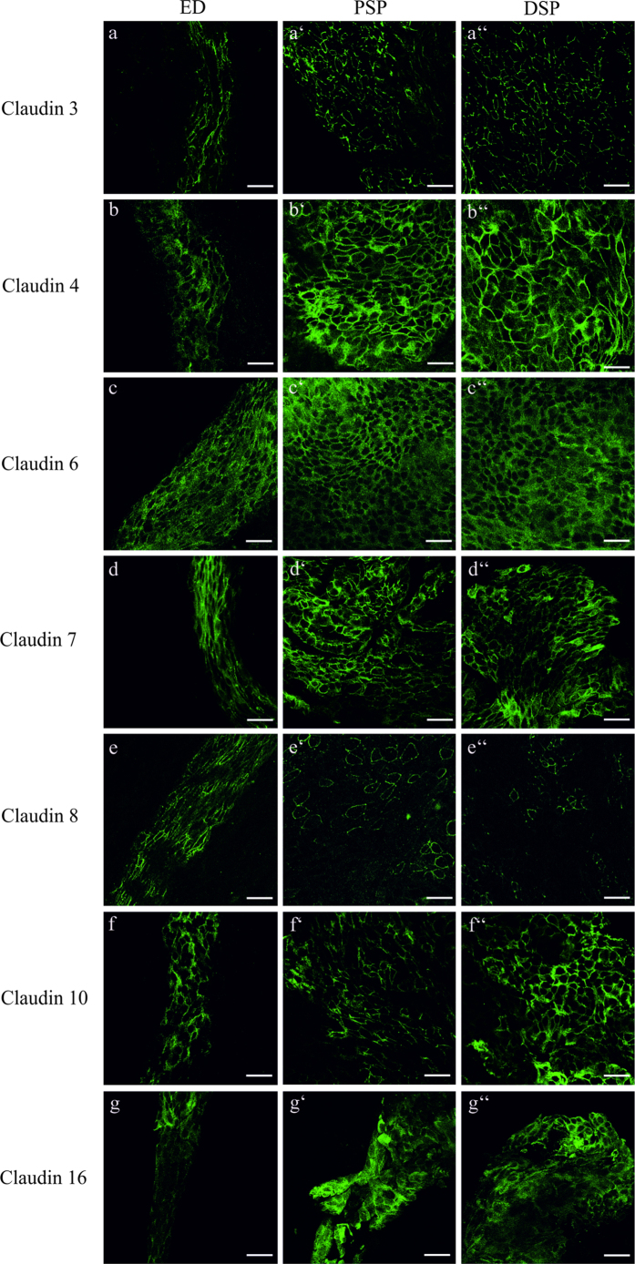
Figure 4. Cellular expression of claudin proteins in the ED and ES epithelium of p4 rat.
Whole mount preparations from rat p4 ES with adjacent ED were stained with the indicated claudin subtype antibody followed by Alexa 488-conjungated anti-rabbit antiserum. (a–a”) Claudin 3 was expressed in the membranes of the epithelia cells of the ED and the PSP and DSP of the ES, whereas no differences in the Claudin 3 signal intensity between the different ES portion and the ED could be observed. (b–b”) Claudin 4 and (g–g”) Claudin 16 immunofluorescence signal was weaker in the cytoplasm and membrane of the epithelial cells of the ED than in the cells of the PSP and DSP. (c–c”) Claudin 6 staining yielded a strong cytoplasmic immunofluorescence signal in the epithelial cells of the ED and ES portions. (d–d”) Claudin 7 immunolabeling was observed in the membranes and in a lower intensity in the cytoplasma of the ED and ES epithelium. (e–e’) Claudin 8 expression was strong in the membranes of the epithelial cells of the ED whereas in the PSP only a few cells showed a Claudin 8 membrane expression. (e”) In the DSP a membrane expression of Claudin 8 was only observed in very few cells. (f–f”) Claudin 10 was expressed in the membranes of the ED and ES epithelium. Scale bars: 10 μm.

