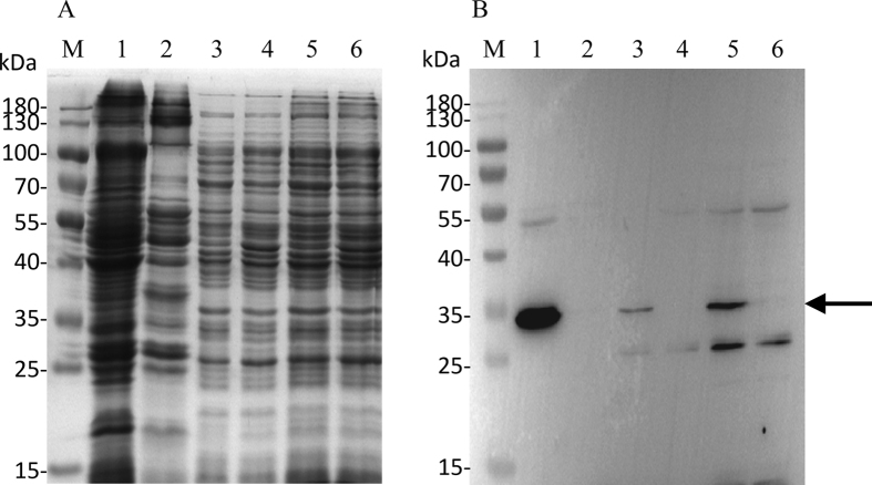
Figure 5. Western blot detection and localization of UPF0118 in Escherichia coli.
For the detection and localization of UPF0118, E. coli KNabc carrying pET-22b-UPF0118 and the empty vector pET-22b (as a negative control) were grown in LBK medium to OD600 between 0.4 and 0.6 at 37 °C, followed by induction by the addition of isopropyl-β-D-thiogalactoside to a final concentration of 1 mM at 28 °C for an additional 6 h and then harvested by centrifugation at 5, 000 g, 4 °C for 10 min and washed three times with Tris-HCl (10 mM Tris -HCl, pH 7.5). The membrane protein fractions, cytoplasmic protein ones and total protein extract from E. coli KNabc/pET-22b-UPF0118 (Lanes 1, 3, 5) and KNabc/pET-22b (Lanes 2, 4, 6) were sampled, respectively, and then used for SDS-PAGE (A) and western blots (B). The position of target protein UPF0118 fused with a N-terminal His6 tag is shown with a solid arrow.

