
Fig. 2.
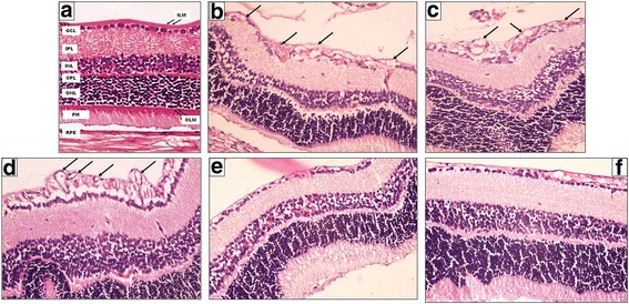
A photomicrograph of a section of a rat’s retina. a Normal arrangement of the different retinal layers in the control negative group (room air). b, c Control positive group (I) and (II) respectively show disorganization in ONL and INL. It also shows the presence of neovascularization both in INL and on the vitreal side of ILM (black arrows). d Another photo of the control positive group (II) shows well apparent neovascularization both in INL and on the vitreal side of ILM (black arrows). e Eye received intravitreal injection of 0.02 ml of 2-Methoxyestradiol (10 μg/ml) shows mild regression of superficial and deep neovessels f Eye received intravitreal injection of 0.02 ml of 2-Methoxyestradiol (100 μg/ml) shows regression of superficial and deep neovessels and preservation of the normal structure of the retina (Hematoxylin-eosin, original magnification (original magnification X400)