
Fig. 4.
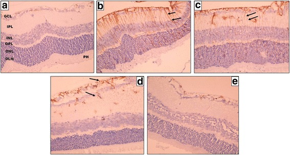
A photomicrograph of a section of a rat’s retina. a Control negative group shows localized positive GFAP immunohistochemical staining confined to GCL, NFL, and ILM. b, c Control positive group (I) and (II) respectively show positive GFAP staining of Muller cells radial processes extended throughout the whole retinal thickness; from the ILM to the OLM (black arrows). d Eye received intravitreal injection of 0.02 ml of 2-Methoxyestradiol (10 μg/ ml) and (e) Eye received intravitreal injection of 0.02 ml of 2-Methoxyestradiol (100 μg/ml) showing limited GFAP expression confined only to GCL, NFL, and ILM (black arrows on) d (original magnification X400)