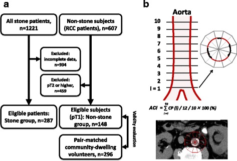
Fig. 1.

Patient selection and measurement of aortic calcification index. Eligible stone patients and non-stone subjects were selected from our database in Oyokyo Kidney Research Institute and Hirosaki University Hospital. In the Stone group, 934 patients were excluded because of incomplete data. The non-stone subjects were selected from renal cell carcinoma (RCC) patients who underwent radical nephrectomy at Hirosaki University Hospital. Of those, we selected early-stage RCC patients (pT1N0M0) for the non-stone control subjects (Non-stone group). Validity of the Non-stone group was evaluated by comparison with pair-matched 296 community-dwelling volunteers from 1166 subjects who participated in the Iwaki Health Promotion Project in 2014 (a). Aortic calcification was quantitatively measured using pretreatment abdominal computed tomography images, scanned 10 times at 10-mm intervals above the abdominal aortic bifurcation. The calcification profile was calculated as the sum of calcification areas of 12 fractions in a single slice divided by 12. The sum of the calcification profile from 10 slices was divided by 10 and multiplied by 100 to obtain the percentage The typical computed tomography (CT) of stone patient is shown. The aortic calcification index (ACI) of this section is 10/12 × 100 = 83.3% (b)
