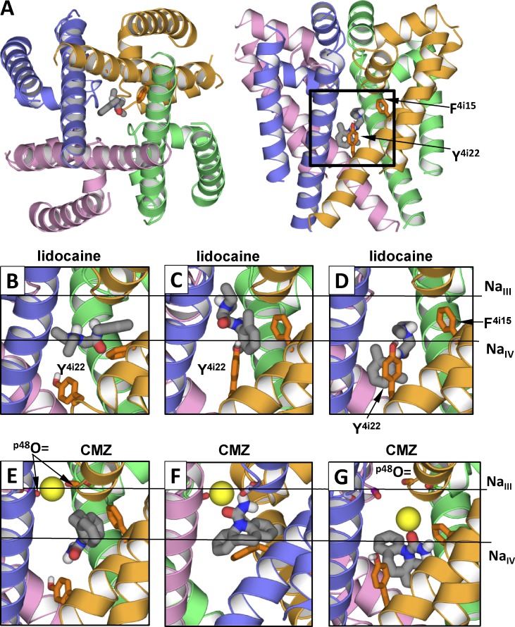
Figure 4.
Examples of low-energy binding modes of lidocaine and CMZ. Side chains of F4i15 and Y4i22 are shown as sticks. Horizontal lines in B–G show levels of site NaIII and putative site NaIV. (A) Location of ligand-binding region. (B–G) Representative structures from the ensembles of low-energy binding modes. (B) Horizontal binding mode of lidocaine. The aromatic group protrudes into the III/IV interface. (C and D) Vertical binding modes with the ammonium group at the levels of NaIII site and NaIV site, respectively. (E) CMZ without tight contacts with NaIII. (F and G) CMZ bound to NaIII. The sodium ion is bound at its innate site seen in the NavMs structure (F) or shifted toward putative site NaIV (G). Note that interactions of lidocaine and CMZ with F4i15 and Y4i22 depend on the ligand-binding modes.

