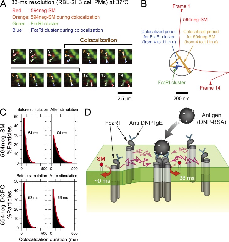
Figure 10.
Single molecules of 594neg-SM were not recruited to FcεRI monomers in quiescent cells, but were recruited to FcεRI clusters induced by multivalent antigens. (A) Typical superimposed videoframe sequences of FcεRI clusters induced by multivalent antigens and a single molecule of 594neg-SM. (B) Trajectories of the FcεRI cluster and the single molecule of 594neg-SM, pointed out by the arrows in A. Colocalization within 240 nm (circle) is shown in the orange (594neg-SM) and blue (FcεRI cluster) trajectories. (C) Duration distributions of the colocalizations of single molecules of 594neg-SM and 594neg-DOPC with FcεRI monomers (before multivalent antigen stimulation) or FcεRI clusters (after stimulation). The exponential colocalization decay time is shown in each box. The number of the observed colocalization events were 212, 241, 224, and 230 from left to right in the top row and then the bottom row. (D) Schematic figure showing transient interactions of SM with FcεRI clusters (after multivalent antigen stimulation) in the PM, but not with FcεRI monomers (before stimulation).

