Figure 6.
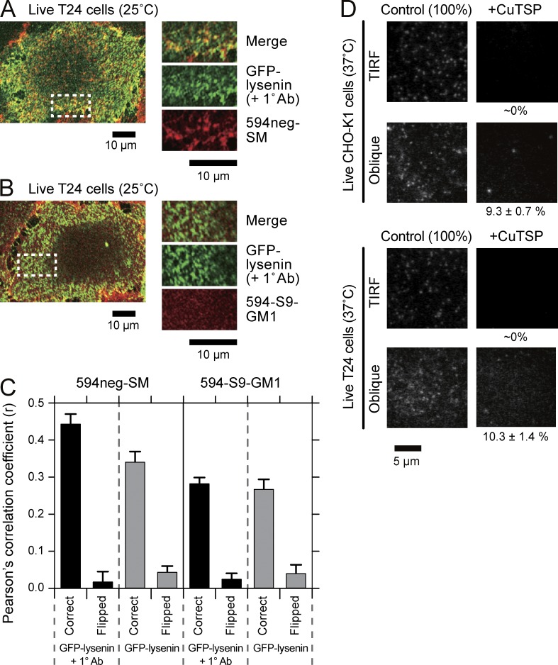
594neg-SM colocalizes with GFP-lysenin at multimolecular levels in PMs and resides in the PM outer leaflet; ∼10% is internalized 15 min after its application. (A and B) Representative confocal fluorescence images of GFP-lysenin and 594neg-SM (A) and 594-S9-GM1 (B) after the application of anti-GFP antibodies to T24 cells. All reactions before the observation were performed at 37°C, and the live cells were observed with a confocal fluorescence microscope at 25°C. (C) Pearson’s correlation coefficients (r) were evaluated for the maximal rectangular regions that could be taken within each cell. Correct, the images of GFP-lysenin and 594neg-SM or 594-S9-GM1 were compared spatially correctly. Flipped, one of the images was rotated by 180° and r was calculated. The difference in r before and after the addition of primary antibodies for 594neg-SM and that without the antibody addition between 594neg-SM and 594-S9-GM1 were statistically significant (P < 0.001). The numbers of images examined were 12 and 11 for 594neg-SM and 13 and 12 for 594-S9-GM1 before and after the addition of primary antibodies, respectively. Error bars indicate standard errors. (D) After incubating T24 and CHO-K1 cells with 594neg-SM for 15 min on the microscope stage, the cells were quickly observed by TIRF and oblique-angle illuminations with a focus near the ventral (basal) PM at single-molecule sensitivities. The membrane-impermeable quencher CuTSP was then added, followed by a second set of observations using TIRF and oblique-angle illuminations (37°C; same view field before and after CuTSP addition). For quantitative data for 594neg-SM, as well as 594neg-DOPC and 594-DOPE, and the method for evaluating percentage molecular fractions in the PM inner leaflet and the cytoplasm, see Table S1 and its notes as well as Materials and methods.

