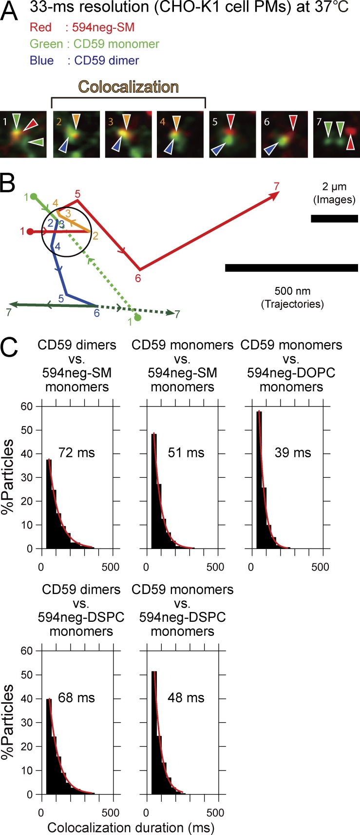
Figure 8.
Single molecules of 594neg-SM were frequently and transiently recruited to CD59 monomers and CD59-homodimer rafts in quiescent cells for 12 and 33 ms, respectively. (A) Typical superimposed videoframe sequences of a 594neg-SM molecule (red spot) and two CD59 molecules (green spots) in the CHO-K1-cell PM, observed at a 33-ms resolution. Two individual CD59 monomers formed a dimer and diffused together (distances ≤240 nm) for the period of four video frames (between frame 2 and frame 6), and simultaneously, these CD59 molecules became colocalized with a 594neg-SM molecule between frame 2 and frame 4. (B) Trajectories of the molecules shown in A. The positions of individual molecules in each image frame shown in A are indicated by the frame numbers. Color coding: 594neg-SM in red (orange during colocalization with CD59); CD59 monomers in green solid and dotted lines, and homo-colocalized CD59 molecules (CD59 homodimer raft) in blue. Colocalization of 594neg-SM and CD59 is shown in the circle, with a 240-nm diameter (see Online supplemental material). (C) Colocalization duration distributions of single molecules of 594neg-SM (+ 594neg-DSPC and 594neg-DOPC) and CD59 monomers or CD59 homodimer rafts. Single-exponential fitting of each histogram provided the colocalization lifetime, which is shown in each box. The numbers of the observed colocalization events were 203, 313, 234, 191, and 288 from left to right in the top row and then the bottom row.

