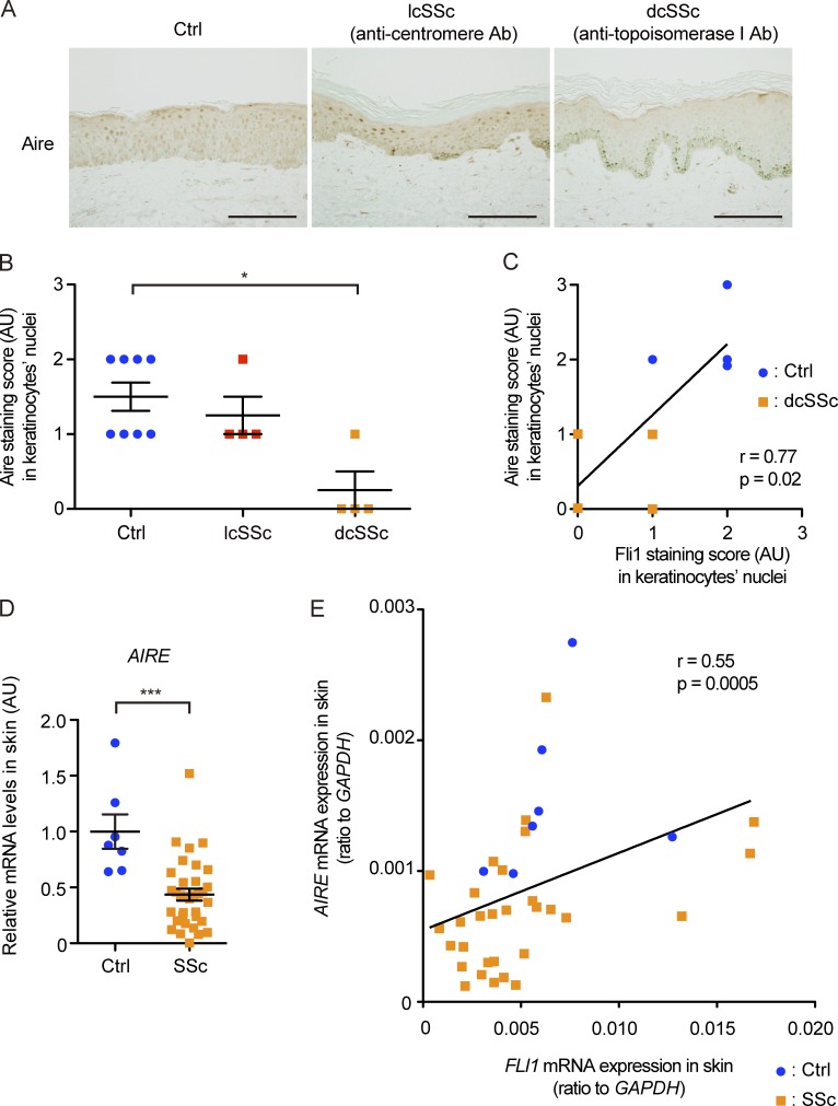
Figure 9.
Aire is expressed in the nuclei of human keratinocytes in vivo, and its expression is significantly down-regulated in SSc patients. (A) Representative images of immunohistochemistry for Aire in healthy controls (n = 8) and lcSSc (n = 4) and dcSSc (n = 4) patients. Bars, 200 µm. (B) The signal intensities of Aire staining in the keratinocytes in A were semiquantitatively evaluated with a four-point grading scale. Kruskal-Wallis test followed by Dunn’s posthoc test was used. (C) Correlation between signal intensities of Aire and Fli1 in the nuclei of keratinocytes in four dcSSc patients and four closely matched healthy controls was analyzed. (D) mRNA expression of the AIRE gene in skin samples from 7 healthy controls and 33 SSc patients was assessed by qRT-PCR. Two-tailed Mann-Whitney U test was used. (E) Correlation analysis of AIRE and FLI1 mRNA expressions in the skin samples used in D (7 healthy controls and 33 SSc patients). (C and E) The solid lines indicate the regression lines. Data are shown as mean ± SEM. *, P < 0.05; ***, P < 0.0001. Ab, antibody; AU, arbitrary units; Ctrl, control.

