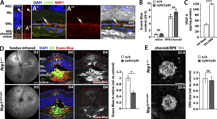
Figure 7.
The NCD promotes vascular leakage, but not neovascularisation, in a mouse model of CNV. (A) Adult eye sections immunostained for NRP1 and CDH5; nuclei were counterstained with DAPI (two independent experiments); NRP1 staining is shown separately on the right. An extension of the squared area is shown at higher magnification in (A’–A’’’); the NRP1 channel is shown separately in (A’’); the DIC image is superimposed in A’’’. RGC, retinal ganglion cell layer; INL, inner nuclear layer; ONL, outer nuclear layer; RPE, retinal pigment epithelium. (B) Evans Blue content in the indicated ocular tissues in Nrp1cyto/cyto mice and wild-type littermates; mean ± SEM; n = 3 mice; ns, P > 0.05; unpaired Student's t test. (C) ELISA shows that VEGF is up-regulated in the RPE/choroid of wild-type mice on D3 after laser injury in the CNV model (n = 4) compared with eyes before laser injury (n = 6); data are expressed as mean ± SEM; the asterisk indicates a significant increase in VEGF levels on D3 (***, P < 0.001; unpaired Student’s t test). (D) Pathological vascular leakage in Nrp1cyto/cyto mice and wild-type littermates. On D3 after laser injury in the CNV model, lesion size was assessed by fundus infrared (IR) imaging (left) before Evans Blue was injected intraperitoneally and dye leakage visualized 24 h later in eye sections counterstained with IB4 and DAPI; the Evans Blue single channel is shown in grayscale on the right hand side. Leakage into the retina at lesion level (as indicated by red) was quantified as the number of Evans Blue–positive pixels integrated for Evans Blue pixel intensity in mutants relative to littermate controls; mean ± SEM; n ≥ 8 mice each; *, P < 0.05 (unpaired Student’s t test). (E) Maximum intensity projections of confocal z-stacks through whole mount RPE/choroids from Nrp1cyto/cyto and wild-type littermates stained for IB4 on D14 after lasering in the CNV model. Quantification of lesion size (right) as number of IB4-positive pixels integrated for IB4-pixel intensity in mutants relative to littermate controls; mean ± SEM; n ≥ 5 eyes each; ns, not significant; P > 0.05 (unpaired Student’s t test). Bars: 25 µm (A); 1 mm (D, left); 200 µm (D, right); 200 µm (E).

