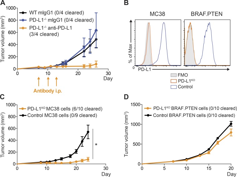
Figure 3.
PD-L1 on MC38 tumor cells is critical for suppression of antitumor immunity. (A) WT or PD-L1−/− mice were given 105 MC38 tumor cells s.c. and treated on days 7, 10, and 13 with anti–PD-1 (339.6A2) or isotype control (mIgG1). Tumors were measured every 2–3 d starting on day 7. (B) Control MC38 and MC38 PD-L1KO (left) or control BRAF.PTEN and BRAF.PTEN PD-L1KO (right) cells were cultured in vitro and stimulated with IFN-γ (20 ng/ml) for 24 h. Expression of PD-L1 was assessed by flow cytometry. (C) WT mice were given either 105 control MC38 or MC38 PD-L1KO tumor cells s.c. and tumors measured every 2–3 d, starting on day 7. (D) WT mice were given either 105 control BRAF.PTEN or PD-L1KO BRAF.PTEN tumor cells s.c., and tumors were measured every 2–3 d starting on day 7. Tumor growth experiments are representative of at least two independent experiments (n = 4–10 mice per group, as indicated). Statistical significance determined by Student’s t test, where P < 0.05 (* indicates significance).

