Figure 2.
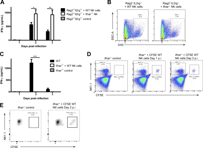
IFNAR is not required directly on NK cells to activate their IFN-γ production. (A) NK cells were isolated from WT or Ifnar−/− spleens and adoptively transferred into Rag2−/−Il2rg−/− mice i.v. 24 h after transfer, mice were infected with 104 pfu HSV-2 ivag, and on days 1–3, vaginal lavages were examined for IFN-γ levels (n = 3; repeated twice with similar results). (B) Spleens were collected on day 3 p.i. and analyzed for DX5 expression. (C) NK cells were isolated from WT spleens, CFSE-labeled, and then adoptively transferred into Ifnar−/− mice. 24 h after transfer, Ifnar−/− mice given WT NK cells, and WT controls were infected with 104 pfu HSV-2 ivag. Day 1–3 p.i. vaginal lavages were examined for IFN-γ content (n = 3; repeated once with similar results). (D) Spleens were examined for CFSE+NK1.1+ adoptively transferred cells on days 1 and 2 p.i. (representative of two independent experiments). (E) Vaginal tissue from Ifnar−/− mice with or without adoptive transfer of CFSE-labeled NK cells was examined on day 2 p.i. for CFSE+NK1.1+ cells (representative of two independent experiments). Vaginal cells were first gated on the CD45+CD3−NK1.1+ population and then examined for CFSE expression. Data in A and C are displayed as mean ± SEM and were analyzed using two-way ANOVA: *, P < 0.05; **, P < 0.01; ***, P < 0.001.

