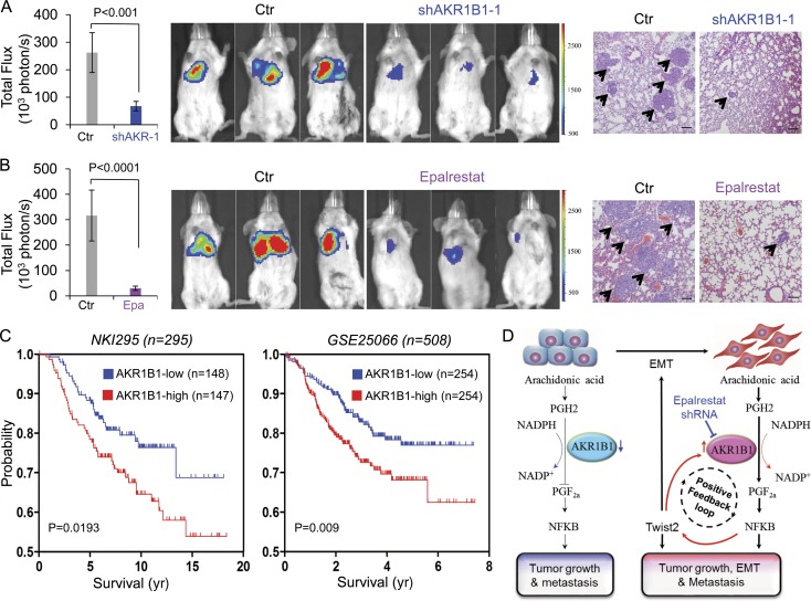
Figure 8.
AKR1B1 promotes metastasis in vivo. (A and B) MDA-MB231 cells with stable empty vector (Ctr) or knockdown of AKR1B1 expression (A) as well as MDA-MB231 cells (B) were injected into SCID mice via the tail vein. For evaluation of epalrestat (Epa), the mice from B then received 50 mg/kg/d epalrestat or sterile water intragastrically. (Left) After 4 wk, the development of lung metastases was recorded using bioluminescence imaging and quantified by measuring photon flux (mean of six animals + SEM). (Middle) Three representative mice from each group are shown. (Right) Mice were also sacrificed. Lung metastatic nodules were examined in paraffin-embedded sections stained with hematoxylin and eosin. The arrowheads indicate lung metastases. Bars, 100 µm. (C) Kaplan-Meier survival analysis of published datasets (NKI295 and GSE25066) for the relationship between AKR1B1 expression and survival time. Differences were performed by the log-rank test. (D) A proposed model to illustrate the regulation of EMT by AKR1B1 through a positive feedback loop, leading to tumorigenicity and metastasis (see Discussion).

