Figure 9.
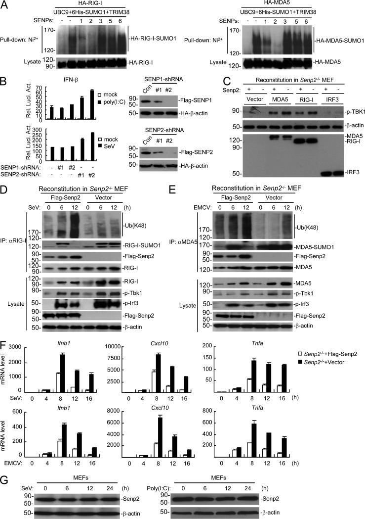
Desumoylation of RIG-I and MDA5 by SENP2 at the late phase of viral infection. (A) Effects of SENPs on sumoylation of RIG-I and MDA5. HEK293 cells were transfected with the indicated plasmids for 24 h before Ni2+ pull-down assays and immunoblotting analysis. (B) Effects of knockdown of SENP1 and SENP2 on SeV- or poly(I:C)-induced activation of the IFN-β promoter. HEK293 cells were transfected with the indicated plasmids for 36 h, and then infected with SeV for 10 h or transfected with poly(I:C) for 18 h before luciferase assays. The knockdown efficiencies of SENP1 and SENP2 shRNAs are shown at the right panels. HEK293T cells were transfected with the indicated plasmids for 24 h followed by immunoblotting analysis. (C) Effects of SENP2 deficiency on RIG-I and MDA5-mediated activation of TBK1. The indicated proteins were transduced into SENP2- and vector-reconstituted SENP2−/− MEFs via retroviral approach, and then cells were harvested, followed by immunoblotting analysis with the indicated antibodies. (D and E) Effects of SENP2 deficiency on sumoylation and K48-linked polyubiquitination of RIG-I and MDA5. Senp2−/− or SENP2-reconstituted MEFs were left uninfected or infected with SeV (D) or EMCV (E) for the indicated times, followed by immunoprecipitation and immunoblotting analysis. (F) Effects of SENP2 deficiency on SeV- and EMCV-induced transcription of downstream antiviral genes. Senp2−/− or SENP2-reconstituted MEFs were left uninfected or infected with SeV or EMCV for the indicated times before qPCR analysis. (G) Effects of viral infection and poly(I:C)-transfected on expression of SENP2. Cells were infected with SeV (left) or transfected with poly(I:C) (right) for the indicated times before lysed for immunoblotting analysis with the indicated antibodies. Data in B and F are from one representative experiment with three technical replicates (mean ± SD). All the experiments were repeated three times.

