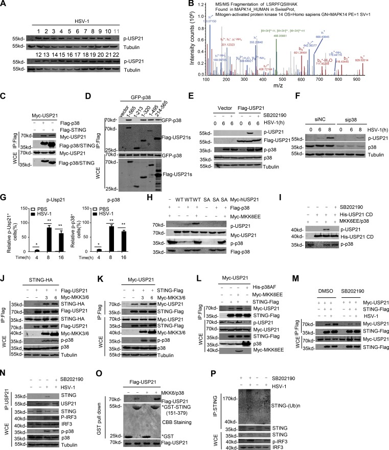
Figure 9.
Phosphorylation of USP21 at Ser538 by p38 MAPK. (A) Screening of kinase inhibitors that affected HSV-1–induced p-USP21 level. The inhibitor details are listed in Table S1. (B) p38 was identified as a USP21 binding partner in the MS assay performed in Fig. 6 A. (C and D) The indicated plasmids were transfected into 293T cells. Cell lysates were then immunoprecipitated with M2 beads and immunoblotted with the indicated antibodies. (E) Immunoblot analysis of phosphorylated and total USP21 in lysates of Flag-USP21 HeLa stable cells treated with DMSO or 10 µM SB202190 and infected for 6 h with HSV-1 (MOI = 1). (F) HeLa were transfected with siNC and sip38, and 48 h later, equal amounts of cells were counted and infected with HSV-1 (MOI = 10) at indicated times. Cell lysate were tested by indicated antibody. (G) FACS analysis of p-USP21 and p-p38 in blood cells obtained from mice (n = 3) that were intravenously injected with HSV-1 (1 × 10⁷ PFU); blood cells were collected at the indicated time. (H) Immunoblot analysis of phosphorylated and total USP21 in lysates of HEK293T cells transfected with indicated plasmid. (I) Immunoblot analysis of phosphorylated and total USP21 with an in vitro kinase assay. Purified recombinant His-tagged USP21 CD was incubated with Flag-tagged p38, which was cotransfected with MKK6EE. (J–L) The indicated plasmids were transfected into 293T cells. Cell lysates were then immunoprecipitated with M2 beads and immunoblotted with the indicated antibodies. (M) The indicated plasmids were transfected into 293T. After 24 h, the cells were pretreated with DMSO or SB202190 for 2 h, after that cells were stimulated with HSV-1 (MOI = 1). Cell lysates were immunoprecipitated with M2 beads and then immunoblotted with the indicated antibodies. (N) Endogenous Usp21 is associated with Sting in L929 cells. L929 cells were pretreated with DMSO or 10 µM SB202190. After 2 h, L929 cells were left untreated or infected with HSV-1 (MOI = 5) for 6 h. Co-IP experiments were performed with anti-USP21, and the immunoprecipitates were analyzed by immunoblotting with the indicated antibodies. (O) Flag-USP21 and Flag-USP21/MKK6EE/p38 were transfected into 293T. The interaction between USP21 and STING was assessed using a GST pull-down assay. All proteins were detected using the indicated antibodies. (P) L929 cells were pretreated with DMSO or with 10 µM SB202190 for 2 h. Next, the cells were infected with HSV-1 or left uninfected for 4 h, and the lysates were subjected to denaturing immunoprecipitation with an anti-STING antibody or normal IgG, followed by immunoblotting with the indicated antibodies. Data from A are screen data from one experiment. Data from B are mass data from one experiment. Data are representative of at least two (C–G and J–P) or three (H and I) independent experiments.

