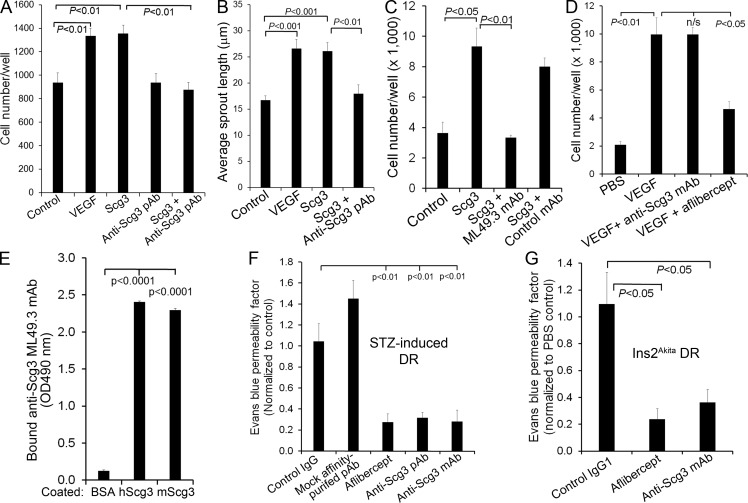
Figure 7.
Anti-Scg3 therapy of DR. (A) Affinity-purified anti-Scg3 pAb blocks Scg3-induced proliferation of HRMVECs. VEGF, 100 ng/ml; Scg3, 1 µg/ml; anti-Scg3 pAb, 2 µg/ml. n = 8 wells. (B) Anti-Scg3 pAb inhibits Scg3-induced spheroid sprouting of HRMVECs. VEGF, 2.5 ng/ml; Scg3, 15 ng/ml; anti-Scg3 pAb, 30 ng/ml. n = 8 spheroids. (C) Anti-Scg3 ML49.3 mAb inhibits Scg3-induced HRMVEC proliferation. Concentrations are as in A. n = 3 wells. (D) Anti-Scg3 mAb cannot neutralize VEGF-induced proliferation of HRMVECs. n = 3 wells. (E) ML49.3 mAb binds to both human Scg3 (hScg3) and mouse Scg3 (mScg3) as detected by ELISA assay. n = 3 wells. (F) Anti-Scg3 therapy of DR in STZ-induced diabetic mice. Anti-Scg3 pAb, mock affinity-purified pAb against an irrelevant antigen, control rabbit IgG, ML49.3 mAb (0.36 µg/1 µl/eye), aflibercept (2 µg/1 µl/eye), or PBS was intravitreally injected. Retinal vascular leakage was quantified by EB assay. Data are normalized to PBS. n = 5 mice (except n = 3 for mock pAb). (G) Anti-Scg3 therapy of DR in Ins2Akita diabetic mice. Doses are as in F. n = 3 mice (4 for anti-Scg3). Experiments were independently repeated three times (A–E) or were repeated twice in a blinded manner (F and G). One representative experiment is shown. Data are ±SEM. One-way ANOVA was used.

