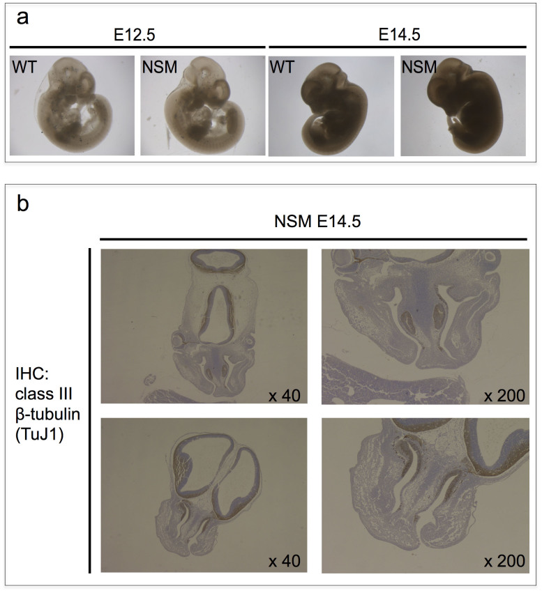
Figure 3. Histological analysis of NSM Tg-positive embryos.
(a) Macroscopic analysis of embryos. There were no apparent abnormalities or difference in size among individual embryos at any stage irrespective of their NSM Tg genetic status. (b) Class III beta-tubulin (TuJ1) immunohistochemical (IHC) analysis of embryonic tissue sections. Representative coronal sections of the embryo heads carrying NSM Tg are shown. Exencephaly, which is frequently associated with lethality in Eker homozygotes, was not observed.

