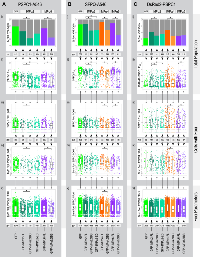
Figure 3. Outcomes of modulating functional IMPα2/α4/α6 levels on paraspeckle marker (endogenous PSPC1/SFPQ or exogenous DsRed2-PSPC1) nuclear transport and paraspeckle localization in HeLa cells.
HeLa cells were transiently transfected with constructs encoding GFP-tagged IMPα2/α4/α6 variants (see Fig. 1A for predicted function) as indicated, with labels at the bottom of each panel and consistent colors used throughout. Paraspeckles were assessed within experimental groups using indirect immunofluorescence with an Alexa Fluor 546 (A546) secondary antibody to detect endogenous PSPC1 (A), using indirect immunofluorescence with an Alexa Fluor 546 (A546) secondary antibody to detect endogenous SFPQ (B) or through exogenous PSPC1 by co-transfecting with a plasmid encoding DsRed2-PSPC1 (C). After analysis pipeline as outlined in Fig. 1B, the primary measures are presented as bar graphs and scatter plots with overlaid box plots indicating the mean and interquartile ranges for each experimental group. Samples with statistically significant differences within IMPα groups are indicated (*). The primary measures include the percentage of foci positive cells (i), the ratio fluorescent signal within the nucleus and cytoplasm (Fn/c) for the paraspeckle marker used (ii), the number of foci detected per cell (iii), the sum foci associated fluorescent signal per cell for the paraspeckle marker used (iv) and the sum foci associated fluorescent signal per foci for the paraspeckle marker used (v). These measures were assessed within groups per entire cell population (i, ii), per foci positive cell populations (iii, iv) or on a per foci basis (v).

