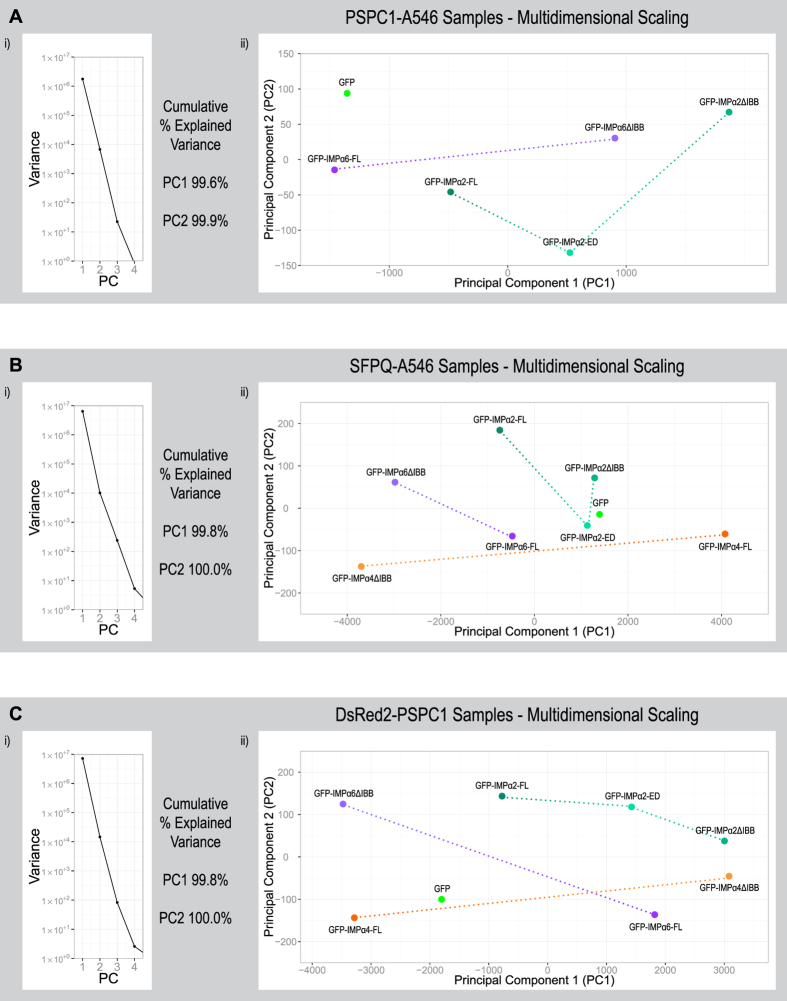
Figure 5. Modulating functional IMPα2/α4/α6 levels affected a plethora measurable paraspeckle related outcomes that can be simultaneously visualised using principal component analysis (PCA).
The results of transiently transfecting HeLa cells with constructs encoding GFP-tagged IMPα2/α4/α6 variants (Tables 1, 2 and 3 and Fig. 2) were used to perform PCA, allowing simultaneous comparisons of multiple parameters and revealing strong patterns between groups. In each experiment PC1 explains >99% of the variance across all parameter and therefore the distances between groups across the X axis (PC1) should be considered as the primary delineator. Paraspeckles were assessed within experimental groups using indirect immunofluorescence with an Alexa Fluor 546 (A546) secondary antibody to detect endogenous PSPC1 (A), using indirect immunofluorescence with an Alexa Fluor 546 (A546) secondary antibody to detect endogenous SFPQ (B) or through exogenous PSPC1 by co-transfecting with a plasmid encoding DsRed2-PSPC1 (C). Parameters used to compare the geometric means of groups within experiments using a specific paraspeckle marker (PSM; A:PSPC1, B:SFPQ, C:DsRed2-PSPC1) were “% cells positive for foci”, “cytoplasmic PSM intensity”, “nuclear PSM intensity”, “PSM Fn/c per cell”, “PSM intensity per cell”, “number of nuclear foci per cell”, “sum volume of nuclear foci per cell”, “sum nuclear foci PSM intensity per cell”, “nuclear foci volume”, “nuclear foci PSM intensity” and “sum nuclear foci PSM intensity”.

