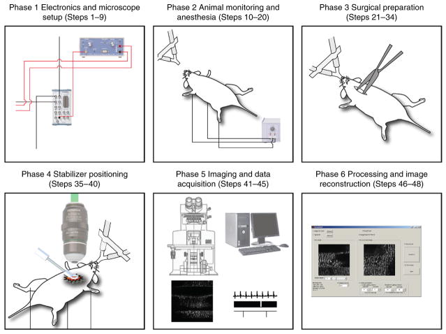
Figure 1.
Schematics of the protocol. This protocol illustrates a methodology for cardiac intravital microscopy in the mouse. The protocol is divided into six different phases. Phase 1, from Steps 1–9, details the electronics and the microscope setup for imaging and data acquisition. Phase 2, from Steps 10–20, illustrates the necessary steps for anesthesia and animal monitoring during the entire duration of the protocol. Phase 3, from Steps 21–34, details the surgical procedure for cardiac intravital microscopy. Phase 4, from Steps 35–40, explains the correct procedure for stabilizer positioning. Phase 5, from Steps 41–45, gives the necessary steps for both microscopy imaging and data acquisition. Finally, Phase 6 from Steps 46–48 highlights data processing routines for final image reconstructions. The approximate cost for the hardware, software and all reagents that are necessary to perform the protocol is in the range of $650,000, $6,000 and $3,000 (in USD), respectively. All animal procedures and protocols were approved by the Institutional Animal Care and Use Committee of the Massachusetts General Hospital, and they are in accordance with the NIH Guide for the Care and Use of Laboratory Animals.

