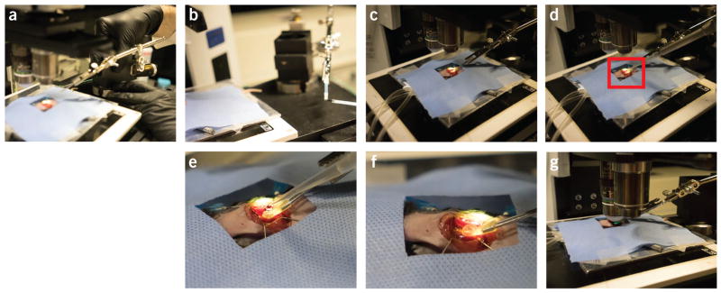
Figure 13.
Positioning of the heart stabilizer. Different phases during the positioning of the stabilizer. (a) Step 35. (b) Step 37. (c) Step 38. (d) Step 39. Water is placed in the stabilizer. (e) Magnified view corresponding to the red box in d. (f) Water fills the stabilizer. (g) Step 40. All animal procedures and protocols were approved by the Institutional Animal Care and Use Committee of the Massachusetts General Hospital, and they are in accordance with the NIH Guide for the Care and Use of Laboratory Animals.

