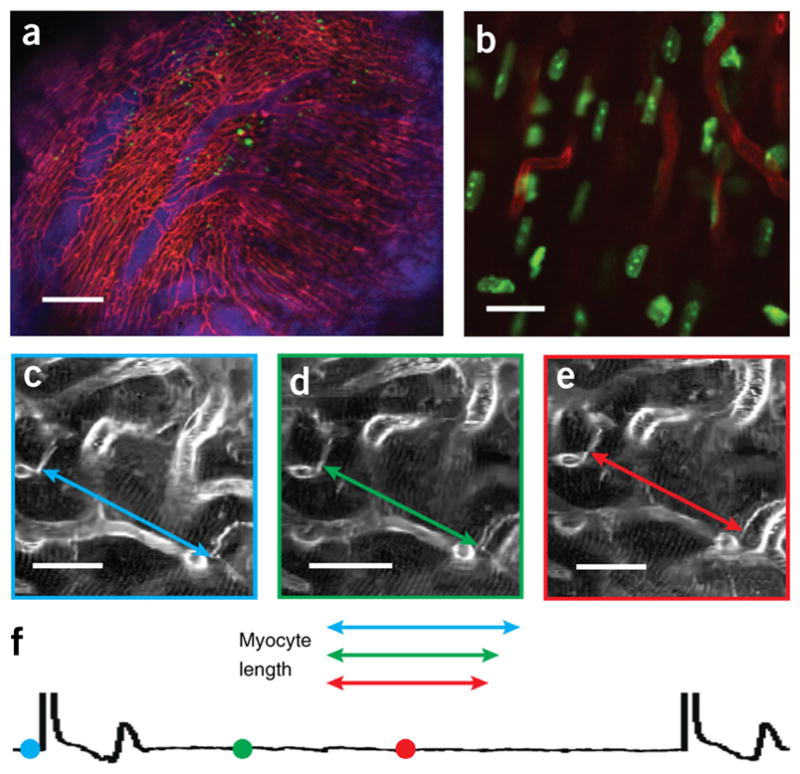
Figure 14.

Intravital imaging of the structure and function in the beating heart at cellular resolution. (a,b) Sequential segmented microscopy with retrospective gating enables multichannel fluorescence confocal microscopy of the heart in diastole over a large field of view (a) and at single-cell resolution (b). Reproduced with permission from ref. 11, Nature Publishing Group. (c–f) Prospective gating allows motion artifact–free imaging of cardiomyocytes at all phases of the cardiac cycle. (c–e) Measurements of the contractile changes of a single cardiomyocyte are obtained at three distinct time points of the cardiac cycle (f). The colored symbols on the cardiac cycle (f) show the stage of the cardiac cycle at which the images shown in c–e were taken. From left to right, the symbols correspond to the images shown in c–e. Scale bars, (a) 200 μm; (b) 10 μm; (c–e) 20 μm. All animal procedures and protocols were approved by the Institutional Animal Care and Use Committee of the Massachusetts General Hospital, and they are in accordance with the NIH Guide for the Care and Use of Laboratory Animals.
