
Figure 4.
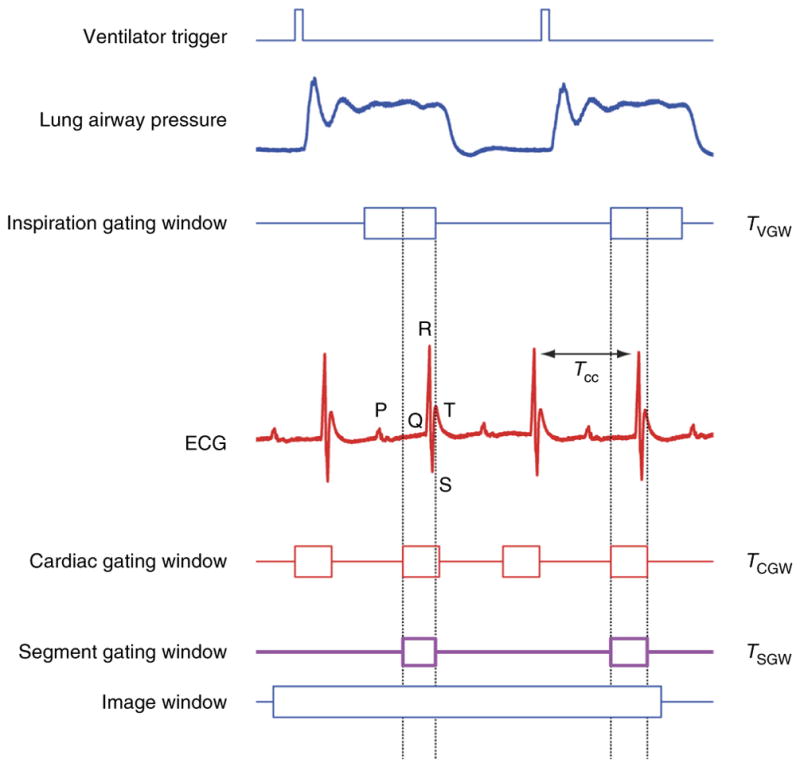
Cardiorespiratory gating. Timing diagram taking into consideration respiratory-induced motion components. The ventilator trigger signal and ECG are recorded and two time gating windows (cardiac TCGW and inspiration TR) are located at points of minimum displacement. TR is usually selected near the end of the inspiration or expiration phase. A new gating window, TSGW, equal to the intersection between the two, needs to be considered in order to isolate within all images all segments that are representative of the same volume.