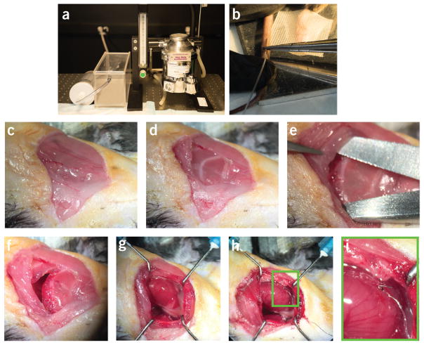
Figure 9.
Highlights from two different phases of the protocol. Important steps during phase 2 (animal monitoring and anesthesia) and phase 3 (surgical preparation) of the protocol are highlighted. (a) Isoflurane gas anesthesia vaporizer and delivery chamber. (b) Insertion of the tail vein i.v. line. (c) Primary skin incision. (d) Cautery through the superficial muscle layer to reveal the ribs. (e) Separation of the fourth intercostal space to access the pleural space. (f) Open thoracotomy incision. (g) Exposed heart with the pericardium intact. (h) Pacemaker lead sutured to the ventricular surface. (i) Expanded view of the area indicated with a green box in h. All animal procedures and protocols were approved by the Institutional Animal Care and Use Committee of the Massachusetts General Hospital, and they are in accordance with the NIH Guide for the Care and Use of Laboratory Animals.

