Fig. S2.
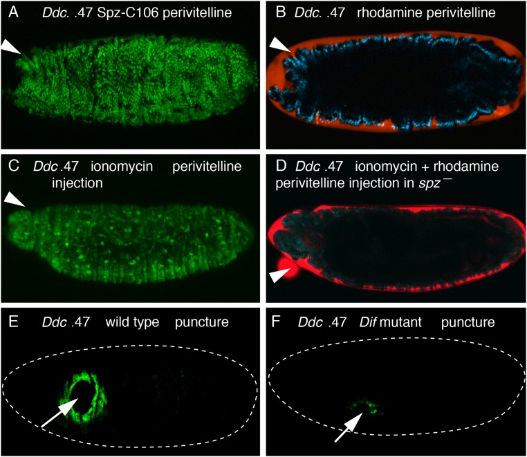
Ddc wound reporter activation by Spz-C106 and ionomycin, and Ddc wound transcription reduction in Dif mutant embryos. (A) Wild-type embryo carrying the Ddc 0.47 wound reporter was injected with Spz-C106 and rhodamine–dextran in the perivitelline space at stage 16, and imaged 5 h later. Global activation of the fluorescent wound reporter protein is seen in epidermal cells. Each green dot is an epidermal nucleus that has accumulated the NLS-fused GFP protein. (B) The same embryo in A was imaged in the rhodamine channel and shows that the red flourescent dye has remained in the perivitelline space atop the apical side of the epidermal cells and has not penetrated into the body cavity. (C) Wild-type embryo carrying the Ddc 0.47 wound reporter was injected with ionomycin in the perivitelline space at stage 16 and imaged 5 h later. Global activation of the fluorescent wound reporter protein is seen in epidermal cells. (D) spz mutant embryo carrying the Ddc 0.47 wound reporter was injected with ionomycin and rhodamine–dextran in the perivitelline space at stage 16 and imaged 5 h later. The signal in the green GFP channel shows no activation of the Ddc wound reporter in the ionomycin-treated spz mutant epidermal cells. The overlaid red signal for rhodamine (red) shows that the red fluorescent dye has remained in the perivitelline space atop the apical side of the epidermal cells and has not penetrated into the body cavity. (E) RNA FISH with a Ddc coding region probe on a wild-type stage 16 embryo 1 h after puncture wounding. Wound induction of Ddc RNA is detected in a normal zone of 5–10 cells distant from the wound edge. (F) The same Ddc probe was hybridized and detected on a Dif mutant embryo that was 1 h postwounding at the time of fixation. Only a few cells have accumulated detectable amounts of Ddc transcripts around wound sites in the spz mutant epidermal cells. See Fig. S3 for quantitation of the effect of Dif mutation on Ddc wound-induced transcription. White arrows mark puncture wound sites, and white arrowheads mark the sites for perivitelline injections. Dashed white lines outline embryos. Anterior is always Left.

