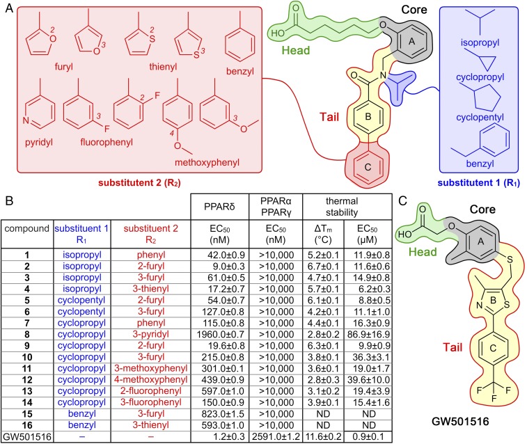
Fig. 1.
Chemical structures of compounds 1–16, GW501516, and their hPPARδ-targeting activity. (A) Structure of compound 1 highlights scaffold design. Regions that occupy arm I (dark-green lines), core (black lines), and arm II (blue or red lines) in the hPPARδ ligand-binding cavity are highlighted. These sectors are accentuated by color fills: head group (light green), core (gray), fixed unit of the tail (yellow), substituent R1 (blue), and substituent R2 (red). (B) Functional groups and associated activities for compounds 1–16 and GW501516. hPPARδ transcriptional responses shown as EC50 values quantified by cell-based luciferase reporter assays. hPPARδ-LBD thermal stabilities shown as both maximal Tm values and EC50 values from fitted dose–response curves (SI Appendix, Fig. S6). ND indicates not determined. (C) Structure of GW501516 with comparable color-coded group designations as in A.

