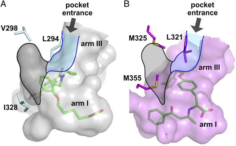
Fig. 4.
Side-chain variants at the hub of the three-arm ligand-binding sites of hPPARα-LBD and hPPARδ-LBD. (A) Accessible surface of the ligand-binding site of 9•hPPARδ-LBD. (B) Accessible surface of agonist-bound hPPARα-LBD (PDB ID code 1K7L) (31). For A and B, the two structures were superimposed to present the three-arm cavities of their ligand-binding sites in the same orientations. Surfaces rendered and colored transparent gray for hPPARδ and transparent purple for hPPARα. Representative protein side chains shown in stick format and labeled hPPARδ (cyan) and hPPARα (magenta) accordingly. Accessible volumes of the central region (black) and arm III (blue) in 9•hPPARδ-LBD are superimposed facilitating comparison with the same sectors in hPPARα. Bound ligands shown in stick format color-coded by atom type, carbon (green), nitrogen (blue), oxygen (red), and sulfur (orange).

