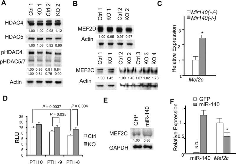
Fig. 3.

Mir140 loss upregulates MEF2C expression and function. (A) Expression or phosphorylation of class II HDACs in primary rib chondrocytes is not affected in Mir140-null mice (KO) compared with Mir140 heterozygous mice (Ctrl). Protein extract isolated from overnight culture of rib chondrocytes (2 mice each genotype) was subjected to Western blot analysis. Signals were quantified by densitometric analysis, normalized by Actin, and expressed relative to Ctrl. (B) MEF2C, but not MEF2D, is upregulated in Mir140-null chondrocytes. Signals were quantified, normalized and expressed relative to Ctrl. (C) Mef2c RNA is upregulated in Mir140-null mice. qRT-PCR was performed using RNA isolated from rib chondrocytes. *p< 0.05, n = 3. (D) MEF2-luciferase assay using primary rib chondrocytes. Cell lysate was prepared for luciferase assay 24 hours after transfection of reporter constructs in the presence of PTH at concentration of 10−9M (PTH-9) and 10−8M (PTH-8). Values of p are indicated. Mir140-null chondrocytes show a modest, but significant, increased MEF2 activity in the presence of PTH. (E, F) Retrovirus-mediated miR-140 overexpression in primary chondrocytes reduces MEF2C expression at the protein (E) and mRNA (F) levels. Primary rib chondrocytes isolated from Mir140−/− mice were infected with viruses expressing miR-140 with GFP (miR-140) or GFP alone (control, GFP). Approximately 60% of cells were infected. Cells were harvested 2 days after infection. Protein expression was quantified, normalized, and expressed relative to control. miR-140 expression was normalized to U6, expressed relative to that of wild-type chondrocytes. ND = not detected. *p < 0.05, n = 3.
