Figure 2.

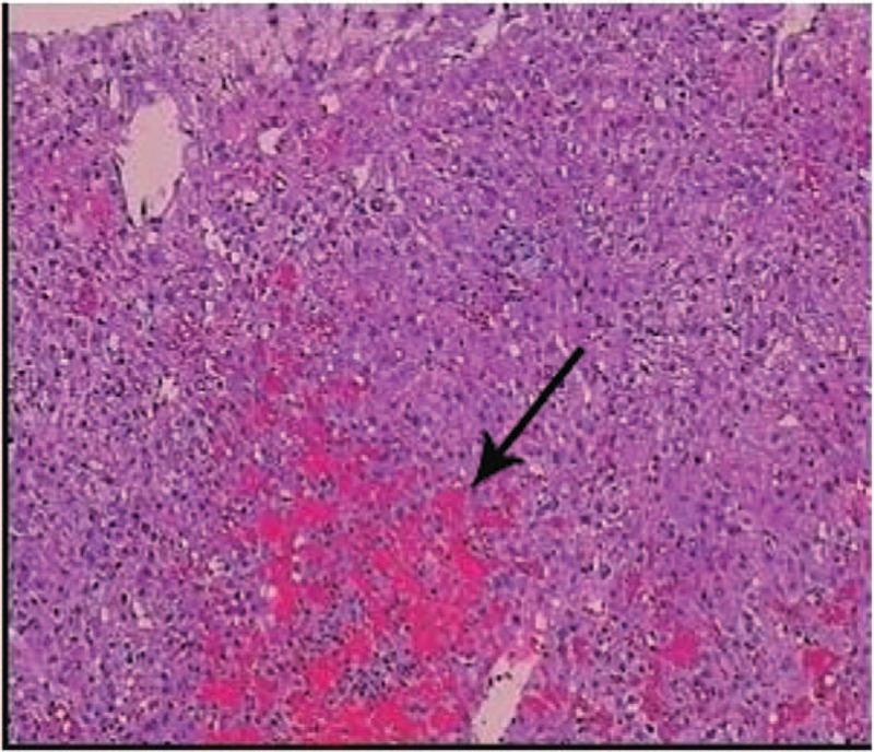
Histology of the liver lesion in case1 indicated a typical feature of peliosis hepatis presenting with multiple sinusoidal dilatations together with blood-filled cystic spaces (arrow). (H&E, x100).

Histology of the liver lesion in case1 indicated a typical feature of peliosis hepatis presenting with multiple sinusoidal dilatations together with blood-filled cystic spaces (arrow). (H&E, x100).