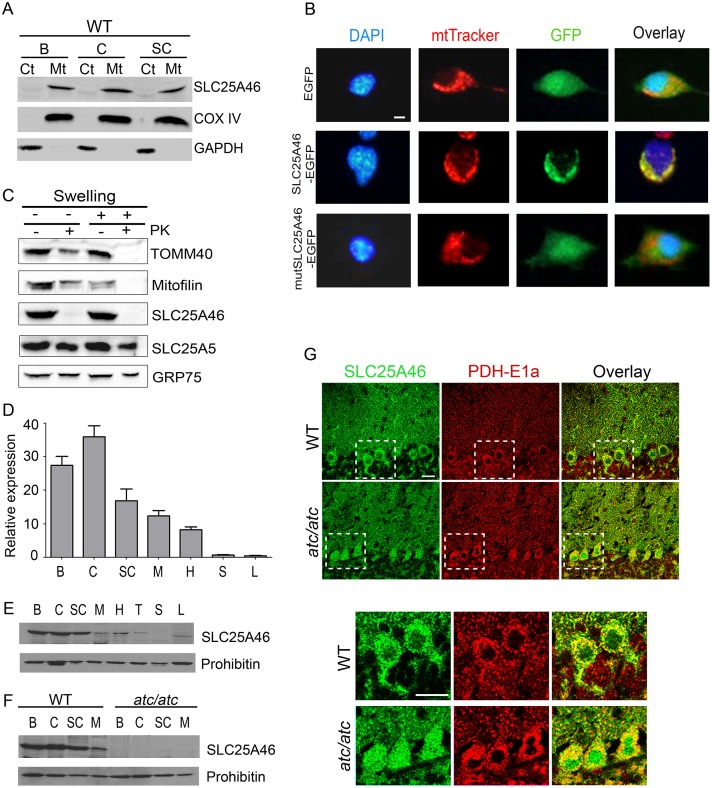
Fig 2. Mitochondrial localization of the SLC25A46 protein.
(A) Cytosolic (Ct) and mitochondrial (Mt) fractions from cerebrum (B), cerebellum (C) and spinal cord (SC) tissues isolated from WT mice were analyzed by immunoblotting with antibodies for SLC25A46, the mitochondrial protein COX IV and the cytosolic protein GAPDH. (B) Fluorescence microscopy of N2A cells transfected with plasmids expressing either EGFP, SLC25A46-EGFP or mutSLC25A46-EGFP carrying the atc mutation. DAPI: nuclear staining, mtTracker: mitotracker for mitochondrial staining. Scale bar: 20μm. (C) Mitochondria and mitoplasts prepared by hypotonic swelling (Sw) of mitochondria from WT cerebellum, were treated with proteinase K (PK) and analyzed by immunoblotting with antibodies against SLC25A46, the outer membrane protein TOMM40, the intermembrane space protein Mitofilin, the integral inner membrane protein SLC25A5 and the matrix protein GRP75. Analysis of the SLC25A46 expression pattern in various tissues from WT mice at the mRNA level with (D) qPCR for mouse Slc25a46 and western blots in mitochondrial extracts from (E) WT mice and (F) WT and atc/atc mice with antibodies against the SLC25A46 and the mitochondrial protein prohibitin. Cerebrum (B), cerebellum (C), spinal cord (SC), muscle (M), heart (H), thymus (T), spleen (S) and liver (L). (G) Immunofluorescence labeling of 4-week-old mid-sagittal cerebellar sections for SLC25A46 shows a clear co-localization with the mitochondrial marker PDH-E1a in PCs of WT mice while a more diffuse distribution is apparent within the PC somata of atc/atc mice indicative of cytoplasmic localization. The insets are shown at higher magnification in the lower panel. Scale bar 20 μm.

