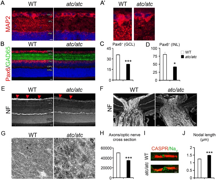
Fig 4. Cellular alterations in the retina and optic nerve of atc/atc mice.
(A) and at higher magnification (A’) Confocal images of 4-week-old mouse retinas showing reduced MAP2 immunostaining in the internal plexiform layer (IPL) of atc/atc mice where the RGC dendrites lie. (B) Amacrine cells immunoreactive for Pax6 and GAD65 are significantly reduced in the atc/atc retina. Quantification of Pax6+ cells in the ganglion cell layer (GCL) (C) and the inner nuclear layer (INL) (D) (n = 3 mice per genotype, p = <0.0001 for the GCL and p = 0.03 for the INL). Confocal images of 4-week-old mouse retina (E) and optic nerve head (F), showing reduced NF immunoreactivity in the innermost part of the retina where RGC axons lie and disorganization of RGC axons in the optic nerve of atc/atc mice. (G) Toluidine blue stained semi-thin optic nerve sections of 4-week-old WT and atc/atc mice. (H) Quantification of axonal numbers per optic nerve section (n = 4 nerves per genotype, p = 0.007). (I) Double immunofluorescence of a myelinated fiber in the optic nerve labeled for CASPR and Nav. Scale bars (A), 10 μm; (B, E, F) 20 μm; (G) 5μm; (I) 1 μm. (J) Quantification of Nav+ nodal length (n = 206 WT and 212 atc/atc nodes pooled from 3 mice/genotype, p<0.00001; two-tailed unpaired Student’s t-test; mean values ± SEM).

