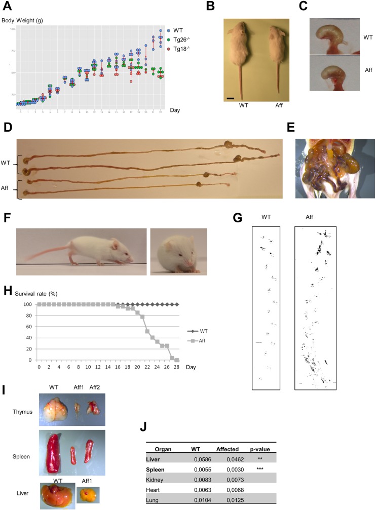
Fig 3. Representative phenotypes in homozygous mice from Tg18 and Tg26 lines (with disruption of the Slc25a46 gene).
(A) Growth curve (birth to 22 days pn) for WT (blue circles), Tg18 homozygous (red circles) and Tg26 homozygous (green circles) mice. Homozygous mice from the two knockout lines stopped gaining weight at ~2 weeks pn. (B) Note size difference between a WT and an affected mouse at 3 weeks post-natal (pn). (C) Representative image of stomach filled with milk for 2 weeks old WT and Tg-/- mice. (D) Gastrointestinal tract (stomach to colon) in WT and affected mice. (E). Representative hemorrhages in the intestinal tract from the oldest affected mice. (F) Representative image of unsteady gait of affected mice. (G) Footprint analyses of WT and Tg-/- mice. The soles of the limbs were labeled with ink. Mice walked on paper in a 10-cm lane surrounded by walls. The ataxic gait is clearly evidenced in the Tg-/- mice, as well as a rapid weakness that prevents them to walk as long as the WT mice. (H) Survival rate curve for WT and Tg-/- animals. Only Tg-/- mice that died prematurely were recorded (27 Tg-/- mice) as well as 40 WT mice. For ethical reasons, most Tg-/- mice were euthanized as soon as they displayed poor health. None of the Tg-/- mice were capable of survival beyond 27 days pn. (I) Thymus, spleen and liver from WT (27 days pn), and affected mice (27 and 22 days pn); note the decreased size of these organs in affected mice. (J) Ratio between organ weight and body weight in 3 weeks old WT (n = 3) and affected mice (n = 4). Liver and spleen were smaller in affected animals. ** p = 0.01, *** p = 0.001, Student test. WT, Wild-Type; Aff, affected; dpn, days post-natal.

