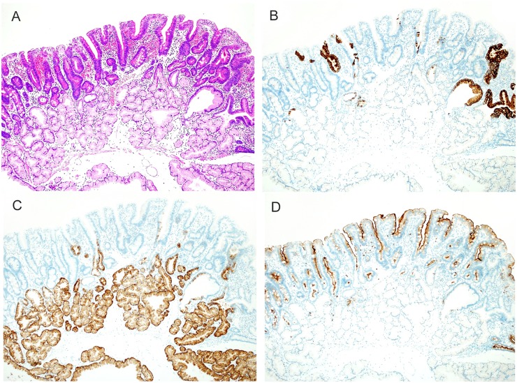
Fig 2. Histologic images of tubular type belonging to intestinal-type.
(A) A hematoxylin and eosin (HE)-stained image. Many tumors were flat and had a tubular structure. (B) Mucin phenotype of tumor (MUC5AC). Among 91 lesions, scattered MUC5AC-positive cells were seen in 70 lesions (76.9%). (C) Mucin phenotype of tumor (MUC6). Same lesion as that shown above in B. Some tumor cells were positive for MUC6. Positivity for MUC6 was also seen in Brunner’s glands. (D) Immunohistochemical findings of tumor (CD10). Most tumor cells, excluding MUC5AC- and MUC6-positive cells, were positive for CD10.

